
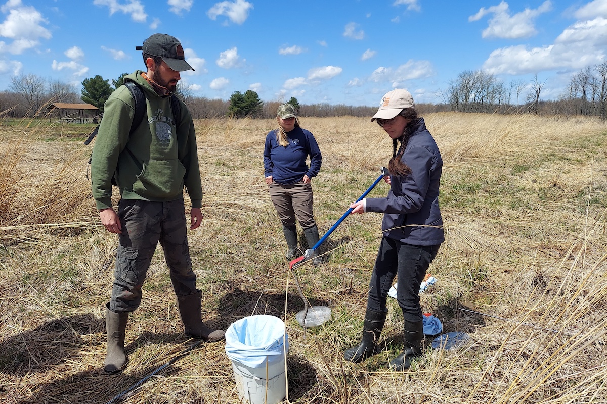
Photo by Courtney Celley, USFWS.

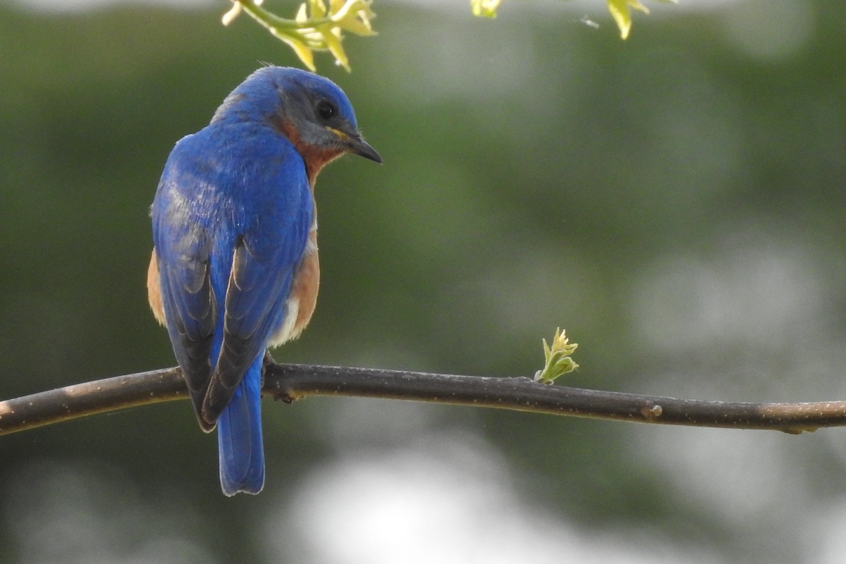
Photo by Courtney Celley, USFWS.
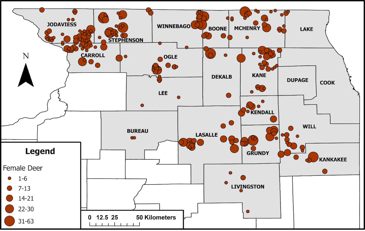
Chronic wasting disease (CWD) is a fatal neurological disease of cervids caused by an infectious protein (prion) that has been detected in Illinois’ wild white-tailed deer herd since 20021. Transmission of CWD is usually through direct contact with an infected animal and its bodily fluids2. Still, consumption of prion-contaminated plants3, water4, or soil5 or vertical transmission from mother to fetus may also cause infection6. That means a fawn could be born infected with CWD, but even if infection does not occur in utero, CWD impacts an animal’s ability to eat, thus a CWD-positive female deer may suffer from nutritional deficiencies7 before the onset of visible signs and the characteristic wasting (weight loss). This raises the question of whether fetal growth is impacted by CWD, particularly the brain and other neurological tissues. To explore this possibility, we used data collected from 18 counties in Illinois (Figure 1) to examine the impact of maternal CWD infection and other variables on fetal head size8, which is directly proportional to brain size9.
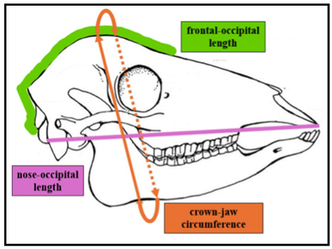
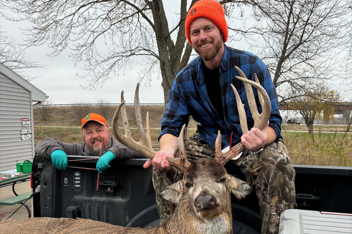
We selected fetuses from known CWD-positive and CWD-negative deer. It should be noted that whether the fetuses were infected was unknown due to difficulties with detecting low prion concentrations at early stages of disease. Three different head measurements were examined in this study: (1) distance between the tip of the nose and the occipital bone, (2) distance from the frontal to the occipital bone, and (3) the circumference of the head from the crown (top) to the jaw (bottom) (Figure 2).
The first step in the analysis was identifying factors that may impact fetal head size, and selecting eight variables: (1) fetal weight, (2) fetal body length, (3) fetal sex, (4) maternal age, (5) maternal weight, (6) number of fetuses in the litter, (7) deer habitat quality and quantity (land cover utility score)10, and (8) the day of the year the female deer died. We also included a ninth variable to account for CWD status. A regression model used the eight variables as the “control variables” to account for differences in head sizes between fetuses (due to fetal, maternal, and environmental characteristics) other than maternal CWD status, isolating the effect of the ninth variable (maternal CWD infection). The results of the model showed that fetal head size increased with fetal body size, and when maternal age and weight increased. Fetal head size also increased with deer habitat quality and quantity, but decreased with larger litters. The model also found that fetuses of CWD-positive female deer had smaller heads than those of CWD-negative deer. Specifically, fetuses of CWD-infected females had nose-occipital lengths and crown-jaw circumferences smaller (6.76% and 11.31% respectively) than their CWD-negative counterparts8.
This finding could have serious implications for fawn survival and success after birth. Though no studies have evaluated the impact of smaller brains on cervids, studies in humans have found delayed and/or impaired cognitive development and problems with vision and hearing11,12. A study of prion diseases in non-human primates suggests that, because smaller brains have less neural tissue, prion-infected animals with smaller brains may die faster13; which may shorten the lifespan of fawns born with CWD or infected soon after birth. This study demonstrates an additional impact of CWD infection, suggesting long-term consequences for the newborn and the herd.
Dr. Jameson Mori is a postdoctoral researcher with the Mateus & Novakofski Chronic Wasting Disease Collaborative Labs. Their research focuses on using data and modeling to determine the impact of chronic wasting disease on white-tailed deer in Illinois and the effectiveness of management efforts to control the disease. They earned their B.S. at the University of Massachusetts Dartmouth and Ph.D. from the University of Illinois Urbana-Champaign.
Dr. Nelda Rivera's research focuses on the ecology and evolution of new and re-emerging infectious diseases and the epidemiology of infectious diseases, disease surveillance, and reservoir hosts’ determination. She is a member of the Wildlife Veterinary Epidemiology Laboratory and the Novakofski & Mateus Chronic Wasting Disease Collaborative Labs. She earned her M.S. at the University of Illinois at Urbana-Champaign and D.V.M at the University of Panamá, Republic of Panamá.
Dr. Nohra Mateus-Pinilla is a veterinary Epidemiologist working in wildlife diseases, conservation, and zoonoses. She studies Chronic Wasting Disease (CWD) transmission and control strategies to protect the free-ranging deer herd’s health. Dr. Mateus works at the Illinois Natural History Survey- University of Illinois. She earned her M.S. and Ph.D. from the University of Illinois Urbana-Champaign.


Submit a question for the author