
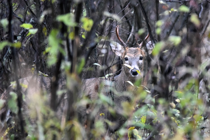
Photo by Jim Hudgins, USFWS.


Photo by Jim Hudgins, USFWS.
Wildlife biologists and natural resource managers know how time and heat can damage the tissues of dead animals. For chronic wasting disease (CWD) testing, there is a ticking clock that starts the moment the cervid dies because tissues degrade rapidly. For years, this left researchers and wildlife managers asking a critical question: will the diagnostic test for CWD still work if the tissue is decomposed?
There are two USDA-approved CWD diagnostic tests: enzyme-linked immunosorbent assay (ELISA) and immunohistochemistry (IHC). In Illinois, both tests are used in sequence to determine the CWD status of the deer; first, ELISA screens for the presence of the infectious protein (prion; Prusiner 1988), then IHC confirms whether the animal is CWD-positive (Varga et al. 2022). Because the prions that cause CWD first accumulate in the lymphatic system, diagnostic testing — a vital part of disease surveillance efforts — uses the retropharyngeal lymph nodes (between the throat and spine) for both ELISA and IHC.
The procedure for lymph node collection from harvested deer followed by the Illinois Department of Natural Resources (IDNR) biologists involves refrigeration until shipment to the diagnostic laboratory to slow down decay and maintain the tissue structure, which is important because there is a difference in the quality/integrity of tissue needed for conducting the two tests. IHC relies on the intact, three-dimensional structure of fresh tissues, making heavily degraded samples difficult or impossible to process. On the other hand, the tissue sample is homogenized prior to ELISA, and so the original tissue structure is not required.
Keeping tissues in cold conditions is easier for samples collected from hunted or sharpshot animals, but in the case of roadkill or other situations where the deer’s carcass has been exposed to the elements for an undetermined length of time, the tissues may already be in advanced stages of decay and lack the structure needed for IHC. This leaves ELISA as the only approved diagnostic testing option, but the limits of ELISA’s ability to detect CWD in decayed lymph nodes was unknown until a study published in 2026 in the journal Prion, titled “Effect of decay on detection of chronic wasting disease (CWD) in lymph nodes using enzyme-linked immunosorbent assay (ELISA),” closed this knowledge gap (Mori et al. 2026). This study involved an experiment that aimed to determine how long CWD prions could be detected in decaying lymph nodes with ELISA. Using white-tailed deer samples from Illinois, we examined how lymph node decomposition for up to 12 months at both refrigeration temperatures (40°F) and warm temperatures (80°F) affected test results.
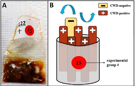
The study included 156 lymph nodes with a known CWD status: 130 CWD-positive and 26 CWD-negative (tissue with an ELISA and IHC test results of negative). We split the samples into groups, each with one negative and five CWD-positive lymph nodes. Each lymph nodes was kept in its own plastic bag with the top open to allow air flow while preventing contact between samples (Figure 1).
Once the groups were assembled, half (n = 13) were placed in a refrigerator (40⁰F) and the other half (n = 13) in an incubator (80⁰F). We then left the groups to decay. After two weeks of decay, and then at every one-month mark, one group was removed from the refrigerator and one from the incubator and immediately frozen. The final groups were removed after 12 months, and all samples were sent back to the diagnostic lab for another round of ELISA testing. Figure 2 shows the percentage of lymph nodes in a group that tested positive for CWD before and after the decay experiment (y-axis) versus the length of the decay period (x-axis). For example, after four months of decay, 5 out of 5 (100 percent) CWD-positive lymph nodes kept their CWD-positive status after refrigeration, whereas only 4 out of 5 (80 percent) of the CWD-positive lymph nodes from the incubator remained positive.
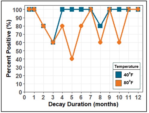
This experiment yielded three main findings. First, it shows that CWD prions can be detected in lymph nodes that have decayed for up to 12 months using ELISA. That means if a lymph node can be located in a carcass, diagnostic testing could still determine the animal’s CWD status. Second, it shows that decay at warmer temperatures results in more initially positive tissues having an undetected ELISA test result at the end of the experiment. It is unclear why this is the case, because the incubator temperature (80⁰F) was far below the recommended temperature for inactivation of prions in contaminated tissue (1832⁰F), but it could be due to some impact of decay on the ELISA test. Third, this study emphasizes the durability of prions and their persistence in cervid tissues. Prions stick around and remain infectious for a long time, to the point where even decay for a full year does not eliminate them, emphasizing the persistence of this pathogen, the importance of testing deer for CWD, and the importance of not moving CWD-positive carcasses or tissues into uninfected areas.
In summary, this study demonstrates that the USDA-approved ELISA test detected CWD in 100 percent of lymph nodes degraded for up to a full year and expands opportunities for surveillance to include older roadkill or degraded field carcasses that were previously assumed to be untestable, as long as the required tissues can be collected.
Dr. Jameson Mori is a research scientist with the Mateus & Novakofski Chronic Wasting Disease Collaborative Labs. Their research focuses on using data and modeling to determine the impact of chronic wasting disease on white-tailed deer in Illinois and the effectiveness of management efforts to control the disease. They earned their B.S. at the University of Massachusetts Dartmouth and Ph.D. from the University of Illinois Urbana-Champaign.
Dr. Nelda Rivera's research focuses on the ecology and evolution of new and re-emerging infectious diseases and the epidemiology of infectious diseases, disease surveillance, and reservoir hosts’ determination. She is a member of the Wildlife Veterinary Epidemiology Laboratory and the Novakofski & Mateus Chronic Wasting Disease Collaborative Labs. She earned her M.S. at the University of Illinois at Urbana-Champaign and D.V.M at the University of Panamá, Republic of Panamá.
Dr. Nohra Mateus-Pinilla is a veterinary Epidemiologist working in wildlife diseases, conservation, and zoonoses. She studies Chronic Wasting Disease (CWD) transmission and control strategies to protect the free-ranging deer herd’s health. Dr. Mateus works at the Illinois Natural History Survey- University of Illinois. She earned her M.S. and Ph.D. from the University of Illinois Urbana-Champaign.





Submit a question for the author